![]()
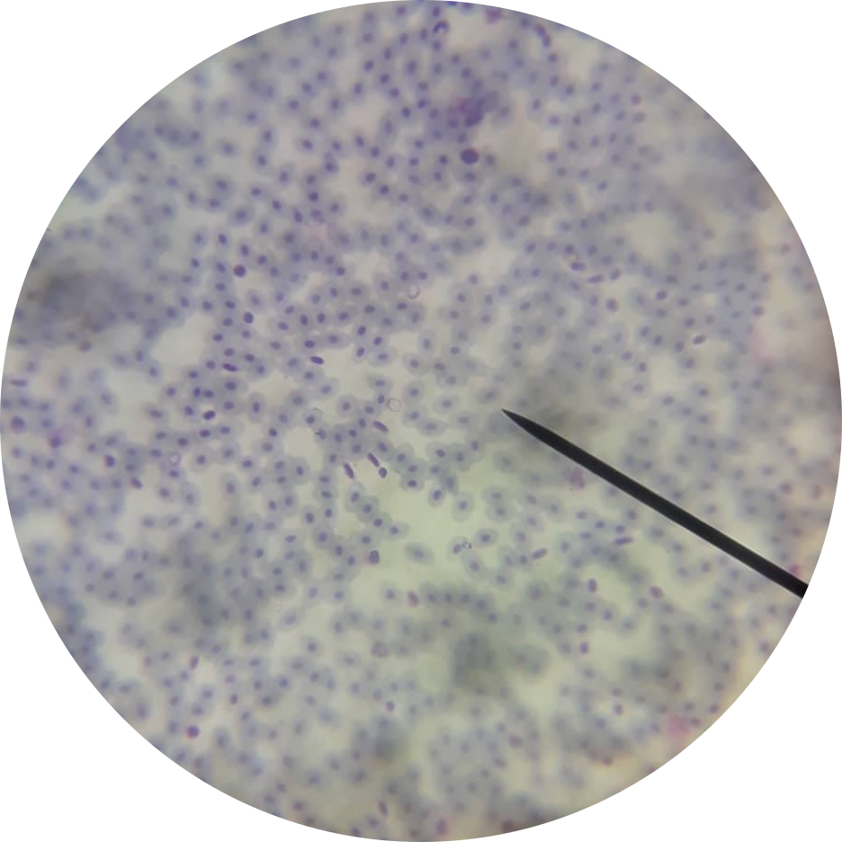
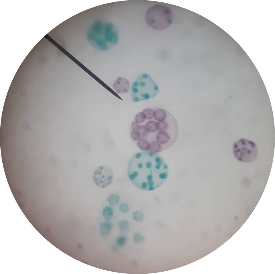
Shown is a bunch of stained blood cells of a frog. The dark middle is the nucleus, while the white part is the cytoplasm.
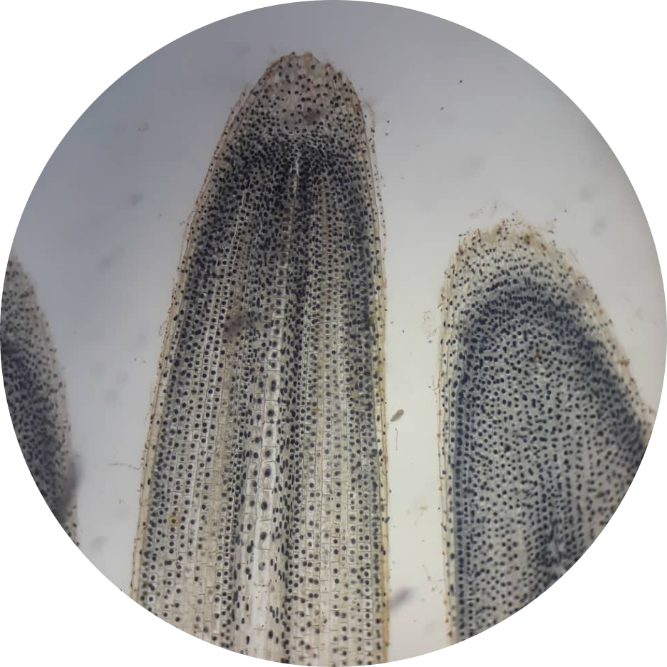
Shown is a root lateral section of an Allium cepa. The darker spots around the tip are the meristematic cells.
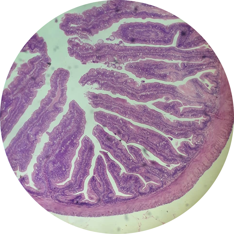
Shown is a stained cross-section of a frog small intestine. The inner, vein-like structure is where the villi is located, while surrounding it is the epithelial tissue. The outer covering is a smooth muscle tissue.
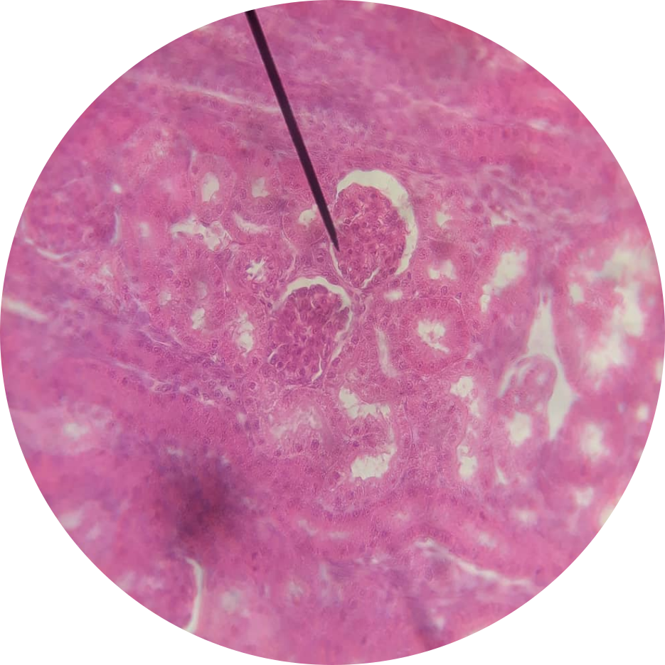
Shown is a cross-section of a kidney. The darker looking parts are the kidney tubules, while right beside these tubules are the cuboidal cells, indicated with a white-colored nucleus.
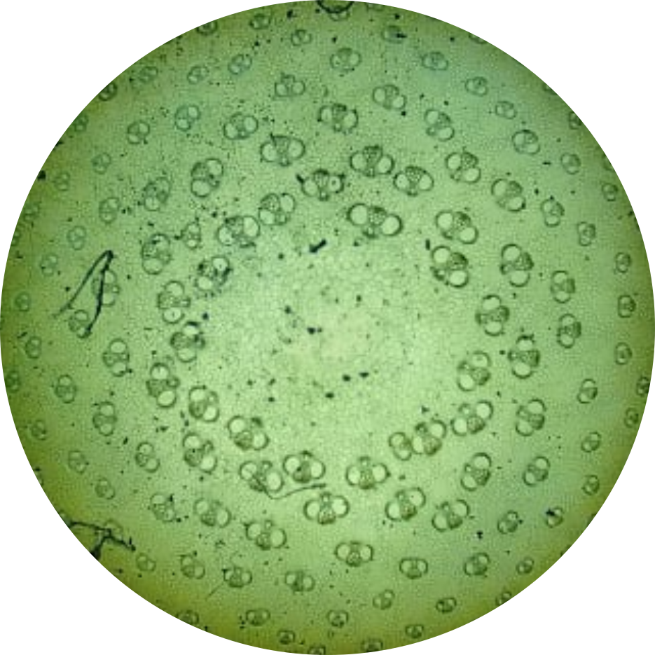
Shown are colonies of the specimen Volvox. Each circle with more cells inside represents one colony.
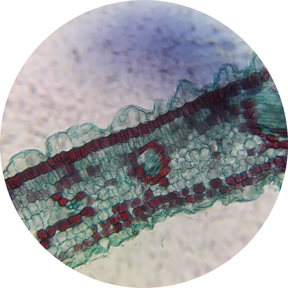
Shown is a cross-section of a stained Ixora leaf. The blue-colored cells are parenchyma cells, and a strand of red-colored cells is one of the veins.
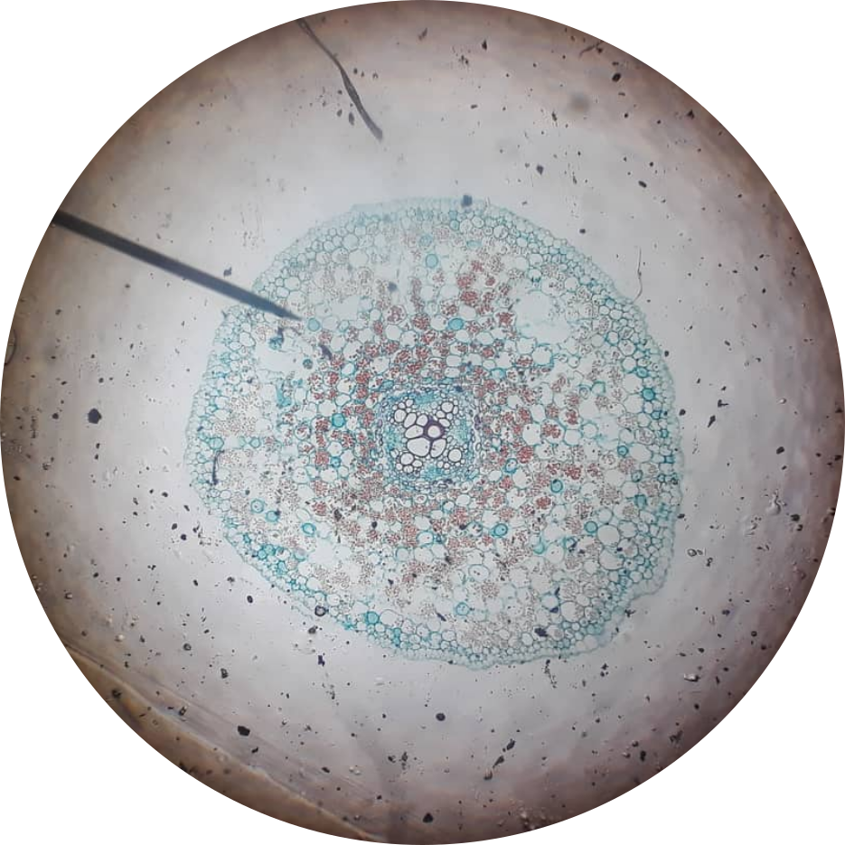
Shown is a root cross-section of a Ranunculus old root. Surrounding the clusters of cells is the dermal tissue; in the very center of this cross-section are the xylem and phloem cells.
Shown is a cross-section of a bamboo stem. The bee-looking cells are composed of two parts: the phloem (the two “wings”), and the xylem (center).